COVID19

Review of literature: introduction and clinical presentations of COVID19

Review of literature: introduction to the COVID19 and clinical presentations

credit: photo by CDC by Unsplash

Virginia Thornley, M.D.
Neurologist
April 2, 2020

Introduction
A new virus emerged in Wuhun, China in December 2019. But the information is still emerging on how to treat it and the exact pathophysiology.

The coronavirus is a type of virus that can infect both animals and humans.
It was named COVID19 for corona virus disease 2019 and renamed SARS-CoV2 which was discovered in the epithelium of the respiratory system of patients from Wuhun, China (1).

COVID-19
COVID19 first occurred December 7, 2019 in the markets of Wuhun, China. The pathogen is the SAR-CoV2. The intermediate host is thought to be the Pangolin. It is a type of RNA virus. The original host is an animal but it jumped to humans. The species pathogen is the B-corona virus. The latency period is about 2-7 days infecting people who have never been exposed to it before(1).
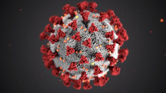
cdc-w9KEokhajKw-unsplash

Clinical presentation
The infection is classified as mild, moderate, severe and critical. Mild cases present with fever, respiratory symptoms and no pneumonia on imaging studies. Moderate is described as those with fever, respiratory symptoms and pneumonia on imaging studies. Severe cases present with respiratory failure with a respiratory rate greater than 30/minute, oxygen saturation or O2 saturation of less than or equal to 93mmHg, PaO2/FiO2 of less than 300mmHg. Critical cases include one of the following: need for mechanical ventilation, shock or organ failure requiring ICU admission. There can be dyspnea leading to acute respiratory distress syndrome (ARDS), metabolic abnormalities that are refractory to correction, shock and thrombosis(3).

Epidemiology
The SARS epidemic which occurred in 2003 affecting China extending to other other Southeast Asian countries, by contrast, lasted 7 months affecting 8096 people resulting in 774 deaths. There was a high mortality rate among hospital personnel of about 21% (1). The COVID19 started December 7, 2019 and is still ongoing at the time of this writing. At the time of this writing, there are 1,040617 affected with 55,188 deaths(2). The numbers continue to climb. It was declared a pandemic by the WHO. Most clinical cases are elderly, however, the coronavirus could be seen in those with diabetes mellitus and hepatitis B. An immunocompromised state is also a risk factor. Male to female ratio based on studies in China is 2.7:1. Mortality rate is 2.1%

References:
1. Xu, J., Zhao, S., Teng, T., Abdalla, A.E., Zhu, W., XIE, L., Wang, Y., Guo, X. Systematic Comparison of Two Animal-to-Human Transmitted Human Coronaviruses: SARS-CoV-2 and SARS-CoV. Viruses. 2020, Feb. 22;12(2).
2. Worldometer, Coronavurus pandemic 2019
3. Feng, Y., Liu, N., Hu, J., Wu, l., Su, G., Zhong, N., Zheng, Z. 4S Respiratory rehabilitation guidelines for patients with pneumonia infected by new Coronavirus. Chinese Journal of Tuberculosis and Respiratory Diseases, 2020, 43: Pre-published online. DOI: 10.3760 / cma.j.issn.1001-0939.2020.0004

Photo by CDC on Unsplash
Neurologybuzz.com

http://virginiathornleymd.com/

unsplash-logoCDC

Standard
PRP

Platelet-rich plasma and mechanism of action

Virginia Thornley, M.D., Neurologist
January 26, 2020

Introduction
Pain is one of the most common conditions that brings a patient to see a physician. Pain is a sign of dysfunction, something is not quite right. There is a plethora of research devoted to understanding the mechanisms.

Some of the more novel approaches are platelet-rich plasma and stem cell therapy. Currently, it is not FDA approved in the United States. It is a novel approach used more extensively outside of the United States.

Mechanism of action
PRP has several growth factors that helps with pain one of which is platelet derived growth factor (PDGF). PDGF arises in the setting of injury when platelets are degranulated. It activates cells which develop high phosphate bonds which leads to specific activities. These activities include mitogenesis, angiogensis and stimulation of macrophage activity. Other growth factors include TGF-beta or transforming growth factor-beta. The target cells are pre-osteoblasts, fibroblasts and marrow stem cells. VEGF or vascular endothelial growth factor is found which stimulates angiogenesis or the formation of new blood vessels. EGF or epidermal growth factor stimulates growth of cells, proliferation and differentiation (1).

Indications
It was found to be helpful in helping injured ligaments and tendons in sports injury.
In one study of 22 patients with intradiscal pain, PRP intradiscal injections were performed which showed encouraging results (2). There are many other indications for PRP in terms of pain control for other conditions.

Summary
PRP shows promising results for various types of pain which was initially used in sports medicine injury but is now expanding to other areas. Large randomized-controlled clinical trials are still needed. However, it is still a viable option. More studies are needed.


References
Jain., N.K., Gulati, M, Platelet-rich plasma: a healing virtuoso, Blood Res. 2016 Mar; 51(1):3-5.
Levi, D., Horn, S., Tyszko, S., Levin, J., Hecht-Leavitt, C., Walko, E., Intradiscal platelet-rich plasma injection for chronic discogenic low back pain: preliminary results from a prospective trial, Pain Med. 2016, Jun; 17(6):1010-1022.

Standard
neurology

Can anti-psychotic agents reduce brain volume?

Virginia Thornley, M.D.
Neurologist, Epileptologist
October 9, 2019
Can medications cause cerebral atrophy? Atrophy refers to shrinkage of the cells causing the appearance of the brain to have less volume than usual.
This question was asked last week. Anti-epileptics such as phenytoin is well-known in the literature and clinically to cause cerebellar atrophy. But what about other agents such as anti-psychotics.
Animal studies
In one animal study, exposure to anti-psychotic drugs showed a reduced volume of brain on volumetric studies. The number of cells remained the same but the volume was increased for cells in the anterior cingulate gyrus which is in the  limbic lobe. The limbic lobe subserves emotions and has influence on memory. Animal studies do not always correlate with human responses.
Human studies
One small study showed that the thalamic volume was reduced after olanzepine administration. This was a small study of 10 patients (2).
While there is some information in the literature the studies are animal studies and small human studies. More information is needed. Based on the current literature, there are not enough significant studies to correlate atrophy with use of anti-psychotics.
References
  1. Vernon, A.C., Crum, W.R., Lerch, J.P., Chege, W., Natesan, S., Modo, M., Cooper, J.D., Williams, S.C., Kapur, S. Reduced cortical volume and elevated astrocyte density in rats chronically treated with anti-psychotic drugs-linking magentic resonance imaging findings to cellular pathology. Biol Psychiatry. 2014, Jun. 15, 75(12):982-90
  2. Khorram, B., Lang, D.J., Kopala, L.C., Vandorpe, RF.A., Rui, Q., Goghari, V.M., Smith, G.N., Honer, W.G. Reduced thalamic volume in patients with chronic schizophrenia after switching from typical anti-psychotic medications to olanzepine. Am J sychiatry. 2006, Nov. 163 (11):2005-7
Standard
dementia

Animal study demonstrating CBD’s (cannabidiol) effects on neuroplasticity and memory loss

Virginia Thornley, M.D.
Neurologist, Epileptologist
August 26, 2019
In an animal study, one group demonstrates that Cannabidiol may help with the neuroplasticity in patients with Alzheimer’s disease (1).
LTP in the hippocampus is the long-term potentiation seen that elevates the efficacy of synapses involved in memory. Beta-amyloid peptide is toxic towards this  feature. When animals were pretreated with CBD the neurotoxicity was found to be reduced against beta-amyloid peptide. The same study showed that it did not involve the 5-HT1a, CB1 or adenosine receptors (1).
There have been other previous studies showing that cannabidiol could have protective effects against the toxic effects of beta-amyloid peptide which is involved in the neurodegenerative process seen in Alzheimer’s disease.
More clinical randomized control trials are needed. Animal studies do not always translate into human studies.
Neurologybuzz.com
References
  1. Hughes, B., Herron, C.E., Cannabidiol reverses deficits in hippocampal LTP in a model of Alzheimer’s disease. Neurochem. Res. 2019, Mar. 44(3):703-713

This is medical information not medical advice. Please consult with your physician.

Standard
multiple sclerosis, Uncategorized

The impact of immunomodulating agents used in multiple sclerosis on the risk of cancer

Virginia Thornley, M.D., Neurologist, Epileptologist
June 14, 2019
Introduction
Multiple sclerosis is already an illness where the immune system recognizes the nervous system specifically the white matter tracts as foreign and attacks it. The complex cascade of mechanisms make adequate treatment challenging. Many treatments focus on the inflammatory mechanism with little attention on the degenerative mechanism involved.
Presentation of symptoms come in a wide variety depending on the the location of the multiple sclerosis plaque in the brain.
Patients may have concomitant morbidities which may make treatment challenging.
 
Immunomodulating agents and its impact on cancer
Many of the newer treatments for multiple sclerosis work at the level of the immune system through immunosuppression, the newer ones tend to be very potent. With greater efficacy comes greater risks including the risk of cancer.
Some of the newer medications can potentially increase the risk of cancer. Higher risk of cancer was found in many reports to occur with use of cyclophosphamide, azathioprine and mitoxanthrone. Fingolimod, natalizumab and alemtuzamab  can potentially increase the risk of cancer, these agents lack long-term data and work through the immune system. Dimethyl fumarate, terifluonimide, ocrelizumab, daclizumab and cladribine merit mandatory risk management plans to detect cancer before its use.
Reference
  1. Lebrun, C., Rocher, F., Cancer risk in patients with multiple sclerosis: potential impact of disease-modifying drugs. CNS Drugs. 2018, Oct. 32(10):939-949 doi:10.1007/s40263-018-0564-y
Disclaimer: This is medical information only not medical advice. Please consult your physician
Standard
cerebellar ataxia

Genetics of Hereditary Cerebellar Ataxia and Hereditary Spastic Paraplegia

Virginia Thornley, M.D.
Neurologist, Epileptologist
March 18, 2019
Introduction
Cerebellar ataxias are rare disorders, only a few types are treatable. This reviews some of the research regarding the genetics of cerebellar ataxias.
Next generation sequencing is a revolutionary way of DNA sequencing that can sequence an entire genome in one day which previously took 10 years. Clinical applications are still pending (1).
Genetics of hereditary cerebellar ataxias
In one study of 87 patients, the genetics were studies. In the probands meaning the first in a genetic line, triplet repeat testing was done. 58% were male. Genetic variants included ANO10, CACNA1A, SPG7 and DRKCG. The detection rate in probands for the trinucleotide repeat was about 13.8%. Those with variants may have a longer duration of disease and a slower progression of the disorder (2).
 
Genetic testing in hereditary spastic paraparesis
In another study where 306 were genetically tested, next generation sequence testing was performed and different genes were found. These include ATL1 (atlastin 1, SPG3),
PAST (spastin, SPG4),  ITPR1, WASHC5 (SPG8),  KIF1A (SPG30), SPG11 spastacsin), KIF5A (SPG10), CYP27A1, and SETX (3).
There are overlapping genetics and clinical symptoms with spinocerebellar ataxia and amyotrophic lateral sclerosis.
Reference
  1. Behjati, S., Tarpey, P., What is next generation sequencing? Arch Dis Child Educ Pract Ed. 2013 Dec; 98(6)236-238
  2. Kang, C., Liang, C., Ahmad, K.E., Gu, Y., Siow, S.F., Colebatch, J.G., Whyte, S., N, K., Cremer, P.D., Corbett, A.J., Davis, R.L., Roscioloi, T., Cowley, M.J., Park, S.J., Sue, C.M., Kumar, K.R. High Degree of Genetic Hetereogeneity for Hereditary Cerebellar Ataxias in Australia, Cerebellum, 2019, Feb. (1):137-146
  3. Elert-Dobkowska, E., Stepniak, I., Krysa, W., Ziora-Jakutowicz, K., Rakowicz, M., Sobanska, A., Pilch, J., Antczak-Marach, D., Zaremba, J., Sulek, A. Next-generation sequencing reveals the broader variant spectrum of hereditary spastic paraplegia and related phenotypes. Neurogenetic, 2019, Feb, doi:10.1007/s10048-019-00565-6
Standard
multiple sclerosis

Ketogenic diet: can it play a role in treating symptoms of Multiple sclerosis?

Virginia Thornley, M.D., Neurologist, Epileptologist
September 19, 2018
@VThornleyMD
Introduction
Multiple sclerosis has no cure at this current moment. It is unclear what is the exact etiology otherwise there would be a cure. Based on research, genetic and environmental factors play a role. Based on MRI observations, there are inflammatory and degenerative components to the pathogenesis.
 
What is the ketogenic diet and how does it pertain the brain
The ketogenic diet was initially found to be effective in treatment of medically refractory seizures. But the underlying concept might be applied to other diseases as well.
Instead glucose as the energy substrate, ketones are utilized, If the supply of glucose is reduced, the energy source is shifted towards the beta-oxidation of fatty acids into ketone bodies. These ketones become the new source of energy and allows increased ATP formation which is the source of energy in the mitochondria, which is the powerhouse of the cell where energy is formed.
37608_434615273840_1627543_n
 
Different lines of thinking regarding pathogenesis of Multiple Sclerosis
There are lines of thought that Multiple sclerosis can be inflammatory versus neurodegenerative. Because of this many agents are directed towards the autoimmune component of the disease process. It is commonly thought that the autoimmune process results in the neurodegeneration seen on MRI.
As evidenced by the “black holes” seen on MRI after acute attacks, there is evidence there is a neurodegenerative aspect. This other line of thinking suggests that it is a degenerative process that triggers the inflammatory response.
It’s been found  that degenerating axons have abnormal mitochondria.
safe_image
Ketogenic diet and inflammation
In one animal study, it was found that the ketogenic diet reduced inflammatory cytokines after 14 days in animals (2).
 
Ketogenic diet and increased ATP
In one animal model with a control group and a group on ketogenic diet, after 3 weeks it was found that those on the ketogenic diet had a higher ATP/ADP ratio which is speculated to contribute towards neuronal stability.

How can the ketogenic diet help with Multiple Sclerosis?
The ketogenic diet reduces the formation of reactive oxygen species. It preserves ATP production when the mitochondria fails. The thought is that the axons start to degenerate once the mitochondria are dysfunctional (1).
In summary
There are no human clinical studies on ketogenic diet and the improvement of multiple sclerosis. Based on pre-clinical studies, there is indication that ketogenic diet may help improve the ATP stores when the mitochondria becomes dysfunctional which may potentially slow neurodegeneration of axons.
The ketogenic diet might reduce inflammation which is thought to be triggered by a neurodegenerative process in Multiple Sclerosis. However, more studies are needed especially human clinical trials. Currently there is not enough evidence to support this based on the available studies as pre-clinical studies do not always correlate in human trials. More studies are needed.

If you’re burned out reading this article check out  Medicine, The Musical brought to you by colleague Writer, Lyricist, Composer – Michael Ehrenreich, M.D. 

Just helping a colleague promote their dream.

Save the date for Medicine. The Musical
safe_image
 
Reference
  1. Storoni, M., Plant, G. The therapeutic potential of the ketogenic diet in treating progressive multiple sclerosis. Mult. Scler. Int. 2015. doi 10.1155/2015/681289
  2. Dupuis, N., Curatolo, N., Benoist, J.F., Auvin, S., Ketogenic diet exhibits anti-inflammatory properties. Epilepsia, 2015. 56(7):e95-98
  3. DeVivo, D.C., Leckie, M.P., Ferrendell, J.S., McDougal, D.B., Jr. Chronic ketosis and cerebral metabolism. Ann Neurol. 1978, Apr. 394):331-337
Standard
Epilepsy

Dravet Syndrome: morphologic abnormalities, role of precision medicine, novel mechanisms for treatment and treatment options

Virginia Thornley, M.D., Neurologist, Epileptologist
@VThornleyMD

August 13, 2018


Introduction

Dravet syndrome is characterized by developmental delay and intractable predominantly myoclonic seizures related to an abnormality in the SCN1A gene. The SCN1A gene encodes for sodium channel Nav1.1 which is voltage gated. It is one of the most pharmacologically resistant types of epilepsy syndromes.

Functional and morphological studies

One animal study using SCN1a(E1099x/HET mouse model for Dravet syndrome demonstrated early seizures which reached its maximum at post-natal week 4. There were less GABAergic neurons that expressed the Nav1.1 subunit in the dentate gyrus in the Het mice. There was a reduced number of inhibitory inputs travelling to the dentate gyrus cells in the Het mice. There was an increase in transmissions of excitatory impulses. The dentate gyral cells were noted to be abnormal morphologically with less arborization and a greater number of spines(1). This correlated with the abnormal excitation and reduced inhibition.

IMG_6486_preview

Fenfluramine

Fenfluramine has been revisited as a treatment option for Dravet syndrome. It is metabolized into norfenfluramine. Fenfluramine and its metabolite norfenfluramine uncouples the association of sigma 1 receptor from the NR1 subunit of NMDA receptors (glutamate N-methyl-D-aspartate). Fenfluramine has serotonergic activity at the 5HT2AR receptor in addition to the activity at the sigma 1 receptor which reduces convulsive activity. Fenfluramine influences the cannabinoid type 1 receptor uncoupling with NMDARs which allowed greater restriction of the NMDAR actions (2).

Ketogenic diet

Ketogenic diet should not be discounted as a therapeutic option (3). In a study of 52 patients with pharmacoresistent epilepsy, spike and sharp wave complexes were reduced on the electroencephalograms of 26 patients which was significant (p<0.5). After a treatment of 12 weeks, there was a noticeable effective rate if seizure reduction of 42%. Motor, language and cognition was found to be improved in 23 patients, although the degree of improvement was not thought to be significant. Some adverse reactions included digestive problems and elevated liver enzymes.

Precision medicine

Because Dravet syndrome is related to a de novo loss of function mutation, great interest has been generated towards precision medicine. This involves targeting the genetic abnormality with treatments tailored towards a patient’s particular genetic make-up.

In one study using precision medicine, the selective activation of the Nav1.1 through the venom Hm1a restored the inhibitory mechanism of the neurons that are responsible for causing seizures in the mice model for Dravet syndrome (4). This may be a novel target for a therapeutic option using precision medicine in the treatment of Dravet syndrome.

Summary

In summary, while Dravet syndrome continues to be a devastating neurological disorder, there is research in precision medicine and other novel therapeutic options that can pave the way for more studies in this area.


https://neurologybuzz.com/
This is info only not medical advice.

Reference

1. Tsai, M.S., Lee, M.L., Chang, C.Y., Fan, H.H., Yu, I.S., You, J.Y., Chen, C.Y., Chang, F.C., Hsiao, J.H., Khorkova, O., Liou, H.H.,Yanagawa, Y., Lee, L.J., Lin, S.W. Functional and structural deficits of the dentate gyrus network coincide with the emerging spontaneous seizures in an Scn1a mutant Dravet syndrome model during development. Neurobiol Dis 2015, May, 77:35-48
2. Rodriguez-Munoz, Maria, Sanchez-Blasquez, Pilar, Garzon, Javier. Fenfluramine diminishes NMDA receptor-mediated seizures via its mixed activity at serotonin 5HT2A and type 1 sigma receptors. Oncotarget. 2018, May, 9(34):23373-23389
3. Qiong, W., Hua, W., Yu, Y., Mei Zhang, J., Yan Liu, X., Ying Fang, X., Hua Yang, F., Jun Cao, Q., Qi, Ying. Ketogenic diet effects on 52 children with pharmacoresistent epileptic encephalopathy: a clinical prospective study. Brain Behav. 2018, May, 8(5):e00973
4. Richards, K.L., Milligan C.J., Richardson, R.J., Jancovski, N., Grunnet, M., Jacobson, L.H., Undheim, EAB, Mobli, M., Chow, C.Y., Herzig, V., Csoti, A., Panvi, G., Reid, C.A., King, G.F., Petrou, S. Selective Nav1.1 activation rescues Dravet syndrome mice from seiuzres and premature death. Proc. Natl. Acad. Sci. U.S.A. 2018, Aug. pii:201804764

Standard
Cancer research and cannabinoids

Cannabinoids: potential role in the detection and reduction of pancreatic tumor load in pre-clinical studies

Virginia Thornley, M.D., Neurologist, Epileptologist

@VThornleyMD

August 1, 2018

Introduction

Cannabinoids are gaining more recognition in treatment not only of pain, seizures and mood disorder but also in a wide variety of conditions. There have been 3 decades of pre-clinical research studying the mechanisms as it relates to the different organ systems. There has been an exponential increase in cannabinoid research especially in light of the demand by grassroot movements for it availability in treating a wide variety of conditions.

As more and more physicians start to recommend it, more symptoms are coming to light which can be ameliorated with medical cannabis. One of the most sought after answer is the deadliest of diseases which is cancer. This seeks to study the mechanisms by which cannabinoids may play a role in reduction of tumor load.

Studies

There are many studies demonstrating the involvement of the endocannabinoid system in modulating the pathogenesis of tumors.

There are no published human clinical trials using cannabinoids in the treatment of the actual underlying pancreatic cancer. Cannabis is labelled under the schedule 1 classification, with that comes the difficulty with procuring the agent because of the bureaucracy and legal red tape that accompanies it. Regardless, there has been an exponential increase in pre-clinical studies in in vitro and in vivo studies.

Detection of pancreatic duct cancer using a CB2 probe

A study showed that the CB2 receptor is highly expressed in pancreatic duct cancer which seems to correlate with  the aggressiveness of the tumor (1). One study reports on using fluorescence imaging on pancreatic duct cancer using an NIR (near infrared) CB2 receptor targeted probe (2). The study found a high level of expression of CB2 receptors in patient samples with pancreatic cancer compared to normal pancreatic tissue. This is significant because it gives information on a specific target for diagnostic and treatment purposes.

CAA11F12-A957-4FAF-B74D-6C7D2CE6E613

Cannabinoid involvement in autophagy through the AMPK pathway

In one study the cannabinoid receptor ligands were discovered to cause autophagy and activate AMPK in pancreatic cancer.  In previous works by the same authors, cannabinoids were found to increase the radical oxygen species. In another study ROS was found to interact with the mitochondria where ATP is produced. AMP is upregulated instead leading to AMPK production which reduces mTOR1c and leads to an increase in autophagy and reduction of cell growth (3).

Possible therapeutic role of CB1 and CB2 receptor ligands on pancreatic cancer

In another study using pancreatic cancer cell lines Panc1, 2 cannabinoid receptor ligands were applied to study the mechanisms of cannabinoids and its possible anti-tumor effect. Cannabinoid ligands GW405833 and arachidonoyl cyclopropramide. The study showed that the cannabinoid ligands were involved in the down-regulation and up-regulation of proteins associated with regulation of cell growth and their energy metabolism. This could be a potential target for therapeutic approaches in pancreatic cancer (4).

Synergistic responses occur when CBD is combined with radiation

Cannabidiol can augment the tumor killing potential when combined with radiation therapy in pancreatic cancer which was studied under in vitro studies. Synergistic responses were noted when 5 micrograms of CBD was combined with 4Gy of radiation therapy in a clonogenic assay. In the same study using mice, there was increased survival in mice with pancreatic tumor using CBD compared to a  control cohort. When CBD was added with SRB or smart biomaterials (agents which are sensitive to environmental factors that allow delivery of other agents in this case CBD to the tumor cells) the mice survived compared to the control cohort with just CBD application alone. This study demonstrates that CBD in conjunction with radiation therapy enhances the tumor killing properties in the treatment of pancreatic cancer (5).

SRB’s or smart radiotherapy biomaterials allow the insertion of payloads which allow the abscopal effects of radiation therapy thereby boosting its results (6). Abscopal refers to the idea that radiation treatment can affect tumors distant from the area treated.

In summary

While there may be a dearth of human clinical trials using cannabinoids for treatment in pancreatic cancer, the pre-clinical studies demonstrate that the endocannabinoid system may play a potential role in the mechanisms, diagnosis and treatment of pancreatic cancer, one of the deadliest tumors, and should not be discounted. More studies are needed especially human clinical trials.

This is info only not medical advice.

References

1. Carracedo, A., Gironella, M., Lorente, M., Garcia, S., Guzman, M., Velasco, G., Iovanna, J.L. Cannabinoids induce apoptosis of pancreatic tumor cells via endoplasmic reticulum stress-related genes. Cancer Res. 2006, Jul, 66(13):6748-55
2. Guo, X., Ling, X., Du., F., Wang, Q., Huang, W., Wang, Z., Ding, X., Bai, M., Wu, Z. Molecular imaging of pancreatic duct adenocarcinoma using the type 2 cannabinoid targeted near-infrared fluorescent probe. Transl Oncol. 2018, Jul. 11(5):1065-1073
3. Dando, I., Donadelli, M., Costanzo, C., Dalla Pozza, E., D’Alessandro, A., Zolla, L., Palmieri, M. Cannabinoids inhibit energetic metabolism and induce AMPK-dependent autophagy in pancreatic cancer cells. Cell Death Dis. 2013, Jun 13, 4 e664
4. Brandi, J., Dando, I., Palmieri, M., Donadelli, M., Cecconi, D. Comparative proteomic and phosphosproteomic profiling of pancreatic adenocarcinoma treated with CB1 and CB2 agonists. Electrophoresis. 2013, May, 34(9-10):1359-1368
5. Moreau, M., Yasmin-Karim, S., Kunjachan, S., Sinha, N., Gremse, F., Kumar, R., Fan Chow, K., Ngwa, W. Priming the abscopal effect using multifunctional smart radiotherapy biomaterials loaded with immunoadjuvants, Front Oncol 2018, 8:56
6. Yasmin-Karim, S., Moreau, M., Mueller, R., Sinha, N., Dabney, R., Herman, A., Ngwa, W. Enhancing the therapeutic efficacy of cancer treatment with cannabinoids. Front Oncol 2018 Apr 24 (8):114
Standard