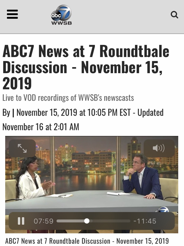
https://www.mysuncoast.com/video/2019/11/16/abc-news-roundtbale-discussion-november/
Neurologybuzz.com

Processed with VSCO with c1 preset

https://www.mysuncoast.com/video/2019/11/16/abc-news-roundtbale-discussion-november/
Neurologybuzz.com
Processed with VSCO with c1 preset

Virginia Thornley, M.D., Neurologist, Epileptologist
@VThornleyMD
August 13, 2018
Introduction
Dravet syndrome is characterized by developmental delay and intractable predominantly myoclonic seizures related to an abnormality in the SCN1A gene. The SCN1A gene encodes for sodium channel Nav1.1 which is voltage gated. It is one of the most pharmacologically resistant types of epilepsy syndromes.
Functional and morphological studies
One animal study using SCN1a(E1099x/HET mouse model for Dravet syndrome demonstrated early seizures which reached its maximum at post-natal week 4. There were less GABAergic neurons that expressed the Nav1.1 subunit in the dentate gyrus in the Het mice. There was a reduced number of inhibitory inputs travelling to the dentate gyrus cells in the Het mice. There was an increase in transmissions of excitatory impulses. The dentate gyral cells were noted to be abnormal morphologically with less arborization and a greater number of spines(1). This correlated with the abnormal excitation and reduced inhibition.

Fenfluramine
Fenfluramine has been revisited as a treatment option for Dravet syndrome. It is metabolized into norfenfluramine. Fenfluramine and its metabolite norfenfluramine uncouples the association of sigma 1 receptor from the NR1 subunit of NMDA receptors (glutamate N-methyl-D-aspartate). Fenfluramine has serotonergic activity at the 5HT2AR receptor in addition to the activity at the sigma 1 receptor which reduces convulsive activity. Fenfluramine influences the cannabinoid type 1 receptor uncoupling with NMDARs which allowed greater restriction of the NMDAR actions (2).
Ketogenic diet
Ketogenic diet should not be discounted as a therapeutic option (3). In a study of 52 patients with pharmacoresistent epilepsy, spike and sharp wave complexes were reduced on the electroencephalograms of 26 patients which was significant (p<0.5). After a treatment of 12 weeks, there was a noticeable effective rate if seizure reduction of 42%. Motor, language and cognition was found to be improved in 23 patients, although the degree of improvement was not thought to be significant. Some adverse reactions included digestive problems and elevated liver enzymes.
Precision medicine
Because Dravet syndrome is related to a de novo loss of function mutation, great interest has been generated towards precision medicine. This involves targeting the genetic abnormality with treatments tailored towards a patient’s particular genetic make-up.
In one study using precision medicine, the selective activation of the Nav1.1 through the venom Hm1a restored the inhibitory mechanism of the neurons that are responsible for causing seizures in the mice model for Dravet syndrome (4). This may be a novel target for a therapeutic option using precision medicine in the treatment of Dravet syndrome.
Summary
In summary, while Dravet syndrome continues to be a devastating neurological disorder, there is research in precision medicine and other novel therapeutic options that can pave the way for more studies in this area.
https://neurologybuzz.com/
This is info only not medical advice.
Reference
1. Tsai, M.S., Lee, M.L., Chang, C.Y., Fan, H.H., Yu, I.S., You, J.Y., Chen, C.Y., Chang, F.C., Hsiao, J.H., Khorkova, O., Liou, H.H.,Yanagawa, Y., Lee, L.J., Lin, S.W. Functional and structural deficits of the dentate gyrus network coincide with the emerging spontaneous seizures in an Scn1a mutant Dravet syndrome model during development. Neurobiol Dis 2015, May, 77:35-48
2. Rodriguez-Munoz, Maria, Sanchez-Blasquez, Pilar, Garzon, Javier. Fenfluramine diminishes NMDA receptor-mediated seizures via its mixed activity at serotonin 5HT2A and type 1 sigma receptors. Oncotarget. 2018, May, 9(34):23373-23389
3. Qiong, W., Hua, W., Yu, Y., Mei Zhang, J., Yan Liu, X., Ying Fang, X., Hua Yang, F., Jun Cao, Q., Qi, Ying. Ketogenic diet effects on 52 children with pharmacoresistent epileptic encephalopathy: a clinical prospective study. Brain Behav. 2018, May, 8(5):e00973
4. Richards, K.L., Milligan C.J., Richardson, R.J., Jancovski, N., Grunnet, M., Jacobson, L.H., Undheim, EAB, Mobli, M., Chow, C.Y., Herzig, V., Csoti, A., Panvi, G., Reid, C.A., King, G.F., Petrou, S. Selective Nav1.1 activation rescues Dravet syndrome mice from seiuzres and premature death. Proc. Natl. Acad. Sci. U.S.A. 2018, Aug. pii:201804764

Virginia Thornley, M.D., Neurologist, Epileptologist
June 3, 2018
Introduction
It is not common for a patient to complain of seizures seeming to increase immediately before a hurricane or a big storm. Do these changes truly correlate with outside environmental factors? This article seeks to review the literature to determine the cause and mechanisms of how weather risk factors might affect epilepsy and frequency of seizures. There is a paucity of information of barometric effects and weather changes on exacerbation of seizure frequency.
Changes in atmospheric pressure correlated with seizures
In one article studying 191 patients, with an increase in atmospheric pressure variability, seizures were noted to increase. The atmospheric pressure was obtained from metropolitan weather stations in Seattle. The maximum, minimum and changes were correlated with the number of seizures being monitored in a telemetry unit over 2005-2006. Patients with known epilepsy had an odds ratio of 2.6 (p=0.02) if the atmospheric pressure varied over 5.5mBar (1).
Higher temperatures correlated with more febrile seizures
In another study of 108,628 pediatric patients from January 2005-December, 2015 were studied regarding the effect of barometric pressure on the frequency of seizures. They were classified as febrile seizures, afebrile, epilepsy or status epilepticus. 53% presented as febrile seizures while 5.9% presented as status epilepticus. Mean atmospheric pressure was 1015.5hPa over the 11 year period. The mean temperature was 14.7 degrees Celsius with a variation of 8.3 degrees Celsius throughout the day. The study demonstrated febrile seizures were influenced by the temperature. At lower temperatures, the emergency room visits were less while at higher temperatures the visits increased (2).

Low barometric pressure, high air humidity increases seizures, high ambient temperature improved seizures
In another study where temperature, barometric pressure, and humidity were correlated with seizure frequency, 604 patients were studied between 2006-2010. The study showed that with a 10.7hPa lower atmospheric pressure there was an increase in seizures by 14%. Those with less severe seizures had an increase of 36%. Relative humidity of >80% correlated with increased seizures of 48%. A high ambient temperature of more than 20 degrees Celsius reduced seizures by 46% (4).
Cold temperature worsen seizures
In a study of 30 patients ages (19-54), patients with epilepsy appeared to have more active seizures during the seasons of spring, autumn and winter and less during summer of about 7%. During stable weather, it was 43% patients and unstable weather 63% had seizures. EEG’s changes occurred more frequently during winter. During winter seizures increased by 40%, in spring it increased 40% and spring by 43.3% (3).
In summary
While anecdotally, there is a correlation of exacerbation of seizure frequency to weather changes, the literature shows mixed results and some of them are small in number. One study showed a correlation of changes of more than 5.5mBar in barometric pressure leading to increased seizures frequency, another showed that it is the reduction in the atmospheric pressure itself that increased seizures. 1 study showed that high humidity may increase seizures. 2 studies showed that cold temperatures worsened seizures, while 1 study showed that higher ambient temperature worsened febrile seizures.
The data that was demonstrated is not uniform in the acquisition of information and there is a large variety of conditions. One study was primarily taken from ER visits another was information from inpatient video EEG monitoring units where the subset of patients may be completely different. In addition, there is a wide heterogeneity in etiologies of seizures which comes into play. Regardless, patients know their own symptoms, usually, if something is noted to trigger an event is it probably real.
Reference

Virginia Thornley, Neurologist, Epileptologist
@VThornleyMD
April 15, 2018
Introduction
The vagal nerve stimulation device is an implanted device that exerts its effort by pulses of electrical activity that stimulates the vagal nerve or cranial nerve X. It had initially been found to work in animal studies in the 1990’s then later applied in clinical studies.
Mechanism of action
For years, the mechanism was unknown and was used rather effectively in the clinical realm. The elucidated mechanisms were thought to be that the vagal nerve stimulator modifies the highly synchronized electrical activity that occurs in epilepsy through desynchronization via the vagal nerve. In addition, there is increased regional cerebral perfusion, and there is increased GABA neurotransmitters which are inhibitory towards electrical activity causing seizures and a decrease in glutamate which is known to increase excitation with the brain. There are GABA-A receptor increases, an increase in locus ceruleus produced noradrenergic substances which are released through the vagal nerve and an increase in serotonin transmissions through the raphe nucleus.
Role in controlling seizures
In the original open-label trial in 5 clinical trials, the vagal nerve stimulation device was found to be effective in reducing seizures by 50%. 454 patients had the implanted device and clinical information was obtained from 440. A cardiac stimulation device was implanted along with a coil in the ipsilateral vagal nerve. At 1 year of implantation, more than 50% of reduction of seizures occurred in 36.8% of patients at year 1, 43.2% year 2, and 42.7% at year 3. The most common side effect at year 2 was hoarseness of about 9.8% and headache in 4.5% and at 3 years there was shortness of breath in 3% (4).
In one retrospective study from 1997 to 2008, 436 patients were found with implanted vagal nerve stimulation devices from ages 1-76, 220 were women and 216 were men. 33 had poor follow-up and 3 had removal due to infection. The mean frequency of seizures was better at 50% reduction. There was 90% better control on 90 patients, >75% control in 162 patients and 50% control in 255 patients, <50% control in 145 patients. Permanent damage to the vagal nerve happened in 2.8% or 11 patients out of the 400 patients (after the removal of the ones lost to follow-up and infected) (5).
Long-term value of vagal nerve stimulating device, effectiveness after 5 years
There have been many studies reported that it may be effective short-term. But there was one pediatric study that reported success in seizure control in longer than 5 years. In a study of 56 pediatric patients ages 4-17, >9.8% were seizure free after 9 months, 24% after 2 years, 46.4% after 3 years and 54% after 5 years.11 out of the 56 patients became seizure free. After 5 years 62% of the patients had fewer seizures after 5 years.
What happens from diagnosis to implantation to use
A patient is identified as medically refractory, meaning a patient who has already failed 2 or more agents. Once control is failed after 2 anti-epileptic drugs after an adequate dosage and trial, the likelihood of being seizure free becomes significantly less. It is usually applied to patients with partial seizures, the most common being temporal lobe epilepsy. After appropriate identification is done, the patient undergoes a procedure where a cardiac device is implanted under the skin which generates an electrical impulse. A wire or coil is attached to the vagal nerve which reacts to this signal and emits an electrical pulse which inhibits the seizure which is electrical activity in the brain by disrupting this through various mechanisms. The device can be programmed to have a set frequency, amount of power and can be set to automatic with features where the patient can apply a magnet to inhibit the seizure when it is about to occur. The magnet is typically swiped over the cardiac device which was implanted over the left side of the chest. The settings can be changed in the doctor’s office adjusting according to the number and frequency of seizures.
Common side effects
Some of the most common side effects reported include hoarseness, cough, throat irritation, dyspnea, insomnia, dyspepsia, and vomiting. The symptoms are related to the location of the device near the nerve causing local irritation and likely due to the functions subserved by the vagal nerve.
Incidental weight loss effect
Vagal nerve stimulation device was applied to treatment-resistant patients with depression where an incidental effect on weight loss was found. One study in 33 patients showed that the vagal nerve stimulator implanted in patients seemed to alter cravings for sweet food which may play a part in weight loss (2). There have been some conflicting studies proving that there is no weight loss in vagal nerve stimulating device at the settings recommended in epilepsy in 21 patients (3). In a large study of 503 patients from 15 study centers, vagal nerve blockade was applied intrabdominally. 294 patients were randomized to treated (192) and to control groups (102). Therapy involved electrical stimulation through an external power source to the vagal nerves in the subdiaphragm which inhibits afferent and efferent vagal transmission. At 12 months, the excess weight loss in the treated group was 17% and in the control group, it was 16%. There was no statistic difference between the two groups, however, the post-study analysis demonstrated a possible result in weight loss related to the system check of the devices using low charges which may have caused weight loss in the control group (6).
In conclusion
There is strong evidence that the vagal nerve stimulation device is effective at reducing seizures of >50% of the medication-resistant epilepsy patient. It is effective even after 5 years of implantation. There are very little side effects which are mild to moderate. In addition, it can cause weight loss.
References:

Virginia Thornley, M.D., Board-Certified Neurologist, Epileptologist
Is marijuana safe for medical use? The take on medical marijuana by the FDA
So far from the FDA official website, the FDA does not recognize medical marijuana coming from the botanical plant with any medical indication. The FDA does not recognize it to be safe or beneficial for any type of disease or condition. The FDA will facilitate any companies interested in bringing quality products including science-based research. The full take of the FDA on marijuana can be found here https://www.fda.gov/NewsEvents/PublicHealthFocus/ucm421168.htm#use
Long-term effects on the brain
Perusing the scientific literature, it is difficult to find any long-term damage to the brain. There was a report in a heavy marijuana user where there was damage to the corpus callosum, possibly worse with young users (1). This is a small study of 11 heavy marijuana users with 11 age-matched cohorts. Diffusion tensor imaging was used. Previous reports alluded towards poor cognition with heavy marijuana use. This study is aligned with that. It was suggested that there may be increased diffusibility within the white matter tracts of the corpus callosum. Young age is thought to make the corpus more susceptible to white matter damage. The only caveat is this is with heavy use and the substance found in recreational marijuana is going to be a different form compared to medical marijuana extracted from the marijuana plant used for medicinal purposes. It is not clear if this report would carry over to medical marijuana users where the preparation of the product is much different(1).

Effect on schizophrenia spectrum diseases
In a large study of 171 patients, it was found that with heavy use of cannabis, the age of onset of schizophrenia spectrum disorders seems to occur earlier (6). This is one of the reasons why in some dispensaries, it is not sold to patients with a history of schizophrenia. There are some anecdotal reports of some patients having a paranoia with medical marijuana that is reversible once taken off.
Effect on the heart, reports of myocardial infarcts and ST elevations
While the literature suggests low toxicity and most side effects are related to cognition and gastrointestinal problems, there are several cannabis-associated myocardial infarcts in the literature. The dispensaries in the state of Florida use a previous history of a previous myocardial infarct as a contraindication in using medical marijuana. These were synthetic drugs used recreationally. There was one case report where a heavy user suffered from an ST elevation and subsequent myocardial infarct after becoming toxic to marijuana used recreationally. In one study, synthetic cannabis was used, the myocardial happened to a young patient where an atheromatous plaque was excluded as the source. Etiology and mechanism are unclear why infarcts should occur. It is quite possible that because it works on the 5HT receptor for anxiety which can cause vasoconstriction, this may be one mechanism. Other studies are needed to elucidate the mechanism of action.
Drug-drug interactions
Because medical marijuana is used as an adjunctive agent for epilepsy, perhaps off-label since it has not been approved through FDA as an anti-epileptic agent yet, it was found that medical marijuana used in conjunction with Clobazam (Onfi) tended to elevate Onfi at higher levels.
In one small clinical study, in 13 patients, 9 had an increase of about 60 in the Clobazam level and by 300 in Norclobazam level. There was, however, a tremendous reduction of seizures by >50% but Onfi (Clobazam and Norclobazam levels) should be monitored (3) on a routine basis to avoid any untoward toxicity.
Other milder symptoms
In one large study on Lennox-Gastaut syndrome where cannabidiol was titrated to a 20mg/kg over a course of 14 weeks, mild to moderate symptoms were noted including pyrexia, sedation, dizziness, and diarrhea. However, the titration rate was very rapid and the patents who were 50kg were quickly at 1000mg within 14 weeks which does not usually happen in the real world. Medications are usually increased over a longer period of time in slower increments.

In summary
While everybody is touting the horn of medical marijuana it is always prudent to stand back and ensure there are no possible risk factors for adverse side effects. The most serious and common seen in the literature appear to be related to schizophrenia spectrum disorders and cannabis associated myocardial infarct. The only caveat is that the literature is peppered with these reports, however, the quality of the recreational drugs are vastly different from medical marijuana which tends to be organic and all natural extracted from the plant in licensed medical dispensaries. The extraction of the medical components is vastly different from the smoked synthetic version of tetrahydrocannabinol. So, is difficult to know if these reports would actually corroborate with use in medical marijuana. The ones with side effects were heavy users of recreational smoked types of marijuana, it is unclear if it was synthetic or organic. As the popularity of medical marijuana progresses, more information will be available regarding the side effect profile.
References